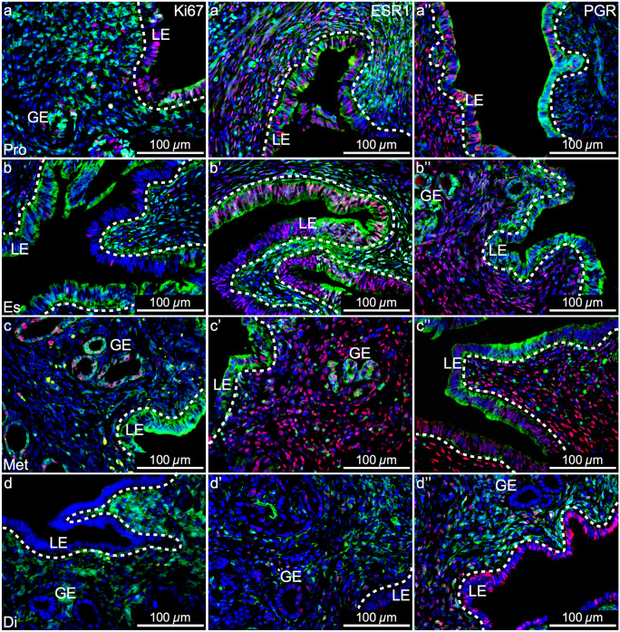
Fig. 7.
Mesenchymal-derived (MD) epithelial cells in adult uteri show characteristics of endometrial epithelial cells but are unique. Representative images of uterine cross sections from adult Amhr2-Cre; Rosa26-EYFP mice in proestrus (Pro) (a–a’’), estrus (Es) (b–b’’), metestrus (Met) (c–c’’), and diestrus (Di) (d–d’’). (a, b, c, d) Ki67 expression by immunofluorescence (IF, red), direct GFP expression in mesenchymal cells an MD-epithelial cells (green), and nuclear DAPI stain (blue). (a’, b’, c’, d’) ESR1 expression by IF (red), direct GFP expression (green), and nuclear DAPI stain (blue). (a’’, b’’, c’’, d’’) PGR expression by IF (red), direct GFP expression (green), and nuclear DAPI stain (blue). ESR1, estrogen receptor alpha; PGR, progesterone receptor; LE, luminal epithelium; GE, glandular epithelium; dotted lines demarcate LE from the underlying stroma

