
Fig. 7.
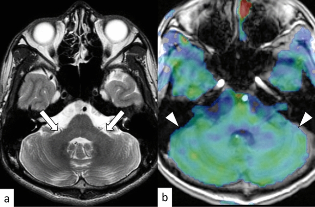
A 21-year-old woman with SCA7. T2-weighted axial image shows mild cerebellar atrophy (a, arrows). Arterial spin labeling image shows decreased blood flow in the cerebellum (b, arrowheads)
Official websites use .gov
A
.gov website belongs to an official
government organization in the United States.
Secure .gov websites use HTTPS
A lock (
) or https:// means you've safely
connected to the .gov website. Share sensitive
information only on official, secure websites.
A 21-year-old woman with SCA7. T2-weighted axial image shows mild cerebellar atrophy (a, arrows). Arterial spin labeling image shows decreased blood flow in the cerebellum (b, arrowheads)