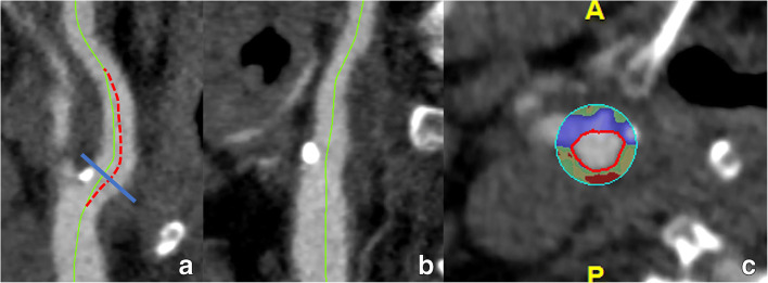
Fig. 10.
The c-MPR-technique and the importance of an adequately positioned centerline. A c-MPR image of the carotid bulb is shown (a, b). The original centerline (green) must be checked in all angles. While it nicely follows the center of the lumen in a frontal view (b), it deviates from it in a sagittal view (a), most notably on the transition from the common carotid artery to the internal carotid artery and more distally in the carotid bulb. As such, it produces in the resulting cross-sectional image an eccentrically positioned lumen (c), which can lead to wrong conclusions. Manual correction (red dotted line in a) is necessary to avoid interpretation mistakes

