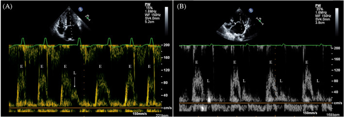
FIGURE 1.

Pulsed wave Doppler mitral inflow showing E waves (E) and L waves (L) in 2 dogs with atrial fibrillation. (A) In this dog with high ventricular rate, only 1 L wave is recognizable during the longer diastolic phase (arrow); in the following cardiac cycle the E and L waves are partially fused. (B) In this dog with lower ventricular rate, L waves are always visible after E waves. Note that all L waves appears as well‐defined positive mid‐diastolic flow velocity with a peak velocity >0.2 m/s
