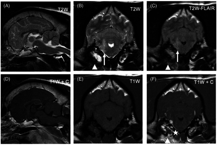
FIGURE 1.

Magnetic resonance images of the head of a dog diagnosed with bacterial meningoencephalitis secondary to otogenic infection (dog 6). From left to right: Top row: sagittal T2W image (A), transverse T2W (B), transverse T2W‐FLAIR (C); Bottom row: sagittal T1W postgadolinium (D), transverse T1W pregadolinium (E), transverse T1W postgadolinium administration (F). Transverse images are at the level of the tympanic bullae and, by convention, the right side of the dog is displayed on the left of the image. There is an ill‐defined T2W and T2W‐FLAIR hyperintensity (compared to normal gray matter) of the right rostral medulla oblongata extending dorsally into the middle cerebellar peduncle (white arrow). The adjacent meninges show contrast enhancement and a focal area of thickening (asterisk). There is bilateral T2W and T2W‐FLAIR hyperintensity of the tympanic bullae contents (more pronounced and homogenous on the right), with bilateral contrast enhancement (again more pronounced on the right). Ventral to the right tympanic bulla there is ill‐defined T2W and T2W‐FLAIR hyperintensity of the soft tissues with marked contrast enhancement (arrowheads)
