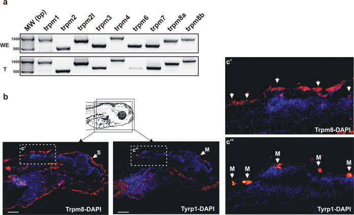
Fig. 3. trpm channel mRNA and TRPM8 protein expression by skin melanophores.
a Representative RT-PCR analysis of mRNA expression for trpm1-trpm8b in the whole embryo and tail at stage 42/43 (N = 3 independent samples). trpm6 mRNA was detected slightly in 1 of 3 independent replicates. b Immunohistochemistry against TRPM8 and Tyrosinase related protein 1 (Tyrp-1) in consecutive sections (schematic). DAPI staining (blue) was used to visualize cell nuclei and facilitate overlapping of adjacent sections. Boxed areas are shown enlarged (c’ and c”). Immunolabel displays co-localization of Tyrp-1 (arrows) in melanophores (M) of the skin (S) with Trpm8. Scale bar = 100 µm.

