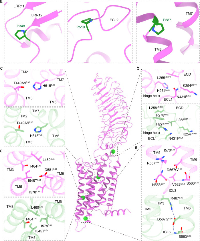
Fig. 6. Detailed structural analysis of mutations in FSHR.
a Three inactive mutations in FSHR. b–e The detailed interactions surrounded residues N431IECL1 (b), T449A/I3.32 (c), I545T5.54 (d) and D567G6.30 (e) mutations. The active FSHR structure is shown in violet. The inactive FSHR structure, shown in light green, was modeled by swiss model based on the highest similar template structure. The four active point mutations are highlighted in green spheres.

