Fig. 4.
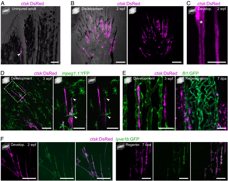
Osteolytic tubules are present in the developing fin, display a tubular morphology, interact with macrophages, align with blood vessels, and are positive for lyve1b:GFP. (A) Confocal image of representative adult uninjured caudal fin displaying ctsk:DsRed+ osteoclasts at the growing edges of rays. (B and C) Confocal images of representative developing caudal fin showing elongated osteoclast structures aligned with fin rays (B) displaying a tubular morphology with a nonfluorescent lumen (asterisk) (C). (D) Confocal images of representative developing pectoral fin, showing direct interaction between macrophages (green) and tubular osteoclasts. Box represents the areas magnified on the Right. (E) Confocal images showing tubular osteoclasts aligning with blood vessels (green) during caudal fin development and regeneration (Left and Right, respectively). (F) Confocal images showing that tubular osteoclasts are positive for lyve1b:GFP, during caudal fin development and regeneration (sets of images on the Left and Right, respectively). Maximum intensity projections in B and D (lower magnification and higher magnification on the Left) and E. Single confocal planes in A, C, and D (higher magnification on the Right) and F. (Scale bars: 50 µm in A and D (higher magnifications), E and F; 100 µm in B and D (lower magnification); 10 µm in C.)

