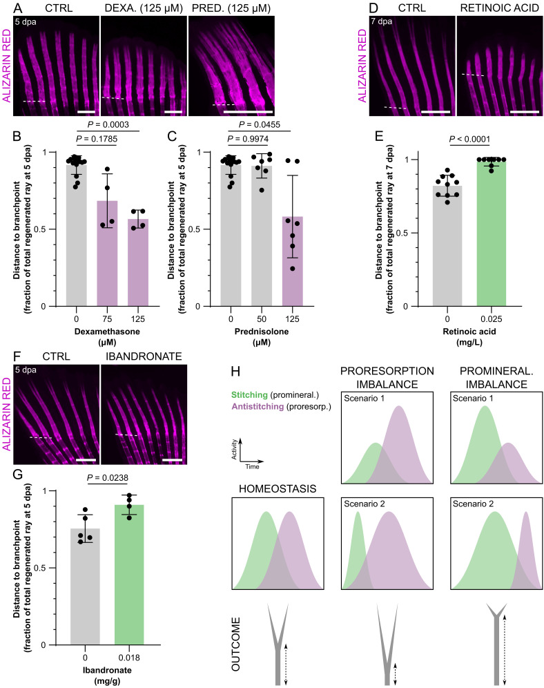
Fig. 6.
Branchpoint position, a proxy of ray bifurcation, is a fast readout of proresorption and promineralization imbalance during bone formation. (A–G) Confocal images (A, D, and F) and quantification (B, C, E, and G) of representative mineralizing rays showing branchpoint positioning upon treatment with dexamethasone (A and B), prednisolone (A and C), retinoic acid (D and E), and ibandronate (F and G). (H) Model depicting how the position of the branchpoints represents proresorption and promineralization imbalance. In scenario 1, the imbalance is due to altered intensity of stitching/antistitching activities; in scenario 2, the imbalance is due to altered duration of stitching/antistitching activities. The dots in the graphs represent individual zebrafish; all graphs show the mean ± SD. Welch one-way ANOVA in B (P = 0.0004) and C (P = 0.0310) and Dunnett T3 post hoc tests for multiple comparisons; two-tailed Student’s t test with Welch’s correction in E; two-tailed Student’s t test in G. (Scale bars: 500 µm.)

