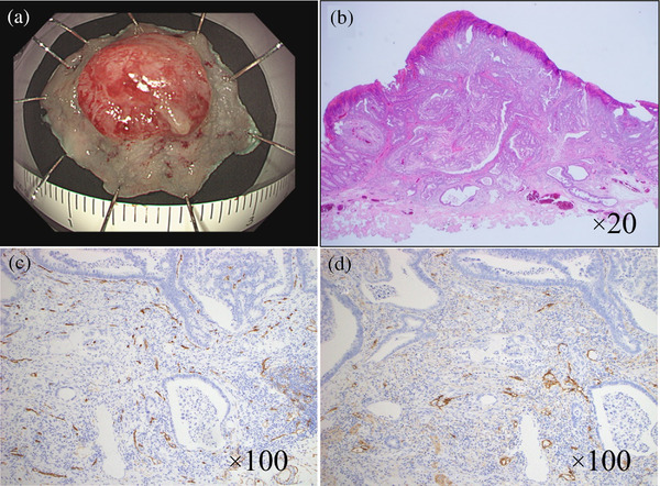
FIGURE 2.

Endoscopic submucosal dissection procedure for the Is+IIc lesion in the sigmoid colon. (a) The tumor was resected en bloc. The resected specimen showed a 0‐I type tumor measuring 9×9 mm. The lateral margin was negative. Microscopic examination revealed a well‐differentiated adenocarcinoma and budding grade 1. The tumor had invaded the submucosa and infiltrated 2000 μm from the surface layer. Immunostaining revealed negative vascular invasion (b–d). (b) Hematoxylin and eosin staining. Magnification of ×20. (c) CD34 staining. Magnification of ×100. (d) D2‐40 staining. Magnification of ×100.
