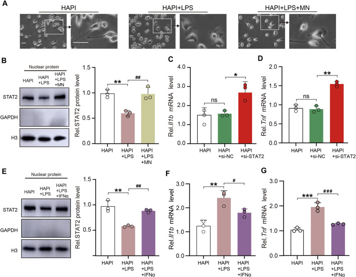
FIGURE 7.
STAT2 in the nuclei negatively regulates pro-inflammatory gene expression in microglia. (A) Representative images of HAPI microglia showed that lipopolysaccharides (LPS) activated microglia and minocycline (MN) significantly inhibited microglia activation (Activated microglia had enlarged cytosomes, shortened protrusions, and amoeba-like cell morphology, scale bar = 50 µm). (B) Nuclear expression of STAT2 was reduced in activated HAPI microglia but it returned to basal level after the treatment of MN (HAPI vs. HAPI + LPS, **p < .01; HAPI + LPS vs. HAPI + LPS + MN, ## p < .01, n = 3, one-way ANOVA followed by Bonferroni’s multiple comparisons test). (C,D) Expression of Il1b and Tnf mRNA was significantly increased in HAPI microglia after transfection of si-STAT2 (n = 3, *p < .05, **p < .01, one-way ANOVA followed by Bonferroni’s multiple comparisons test). (E) Nuclear expression of STAT2 was restored after treatment of IFNα (HAPI vs. HAPI + LPS, **p < .01; HAPI + LPS vs. HAPI + LPS + IFNα, ## p < .01; n = 3, one-way ANOVA followed by Bonferroni’s multiple comparisons test). (F,G) Treatment of IFNα reversed the LPS-induced upregulation of Il1b and Tnf mRNA expression in HAPI microglia (HAPI vs. HAPI + LPS, **p < .01, ***p < .001; HAPI + LPS vs. HAPI + LPS + IFNα, # p < .05, ### p < .001, n = 3, one-way ANOVA followed by Bonferroni’s multiple comparisons test).

