Figure 6.
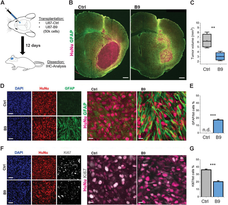
Forced differentiation reduces in vivo tumorigenesis. A, Schematic representation of the transplantation procedure. B, Brain slices from animals transplanted with either non-reprogrammed or reprogrammed U87 cells stained for the human nuclei marker HuNu (magenta) and GFAP (green). Scale bar, 200 μm. C, Volumetric quantification of the tumor mass based on HuNu IHC staining. D, High magnification images of GFAP-HuNu staining from sections shown in B. Scale bars, 50 μm (left) and 10 μm (right). E, Relative population of GFAP-expressing cells in the U87-B9 and control groups. F, High magnification images of Ki67-HuNu staining. Scale bars, 50 μm (left) and 10 μm (right). G, Quantification of the relative number of Ki67-positive cells over the total HuNu-positive cells in the U87-B9 and control groups. n = 5 mice for each experimental condition. **, P < 0.01; ***, P < 0.001. Bar graphs show mean frequency (± SEM). n.d., not detected.

