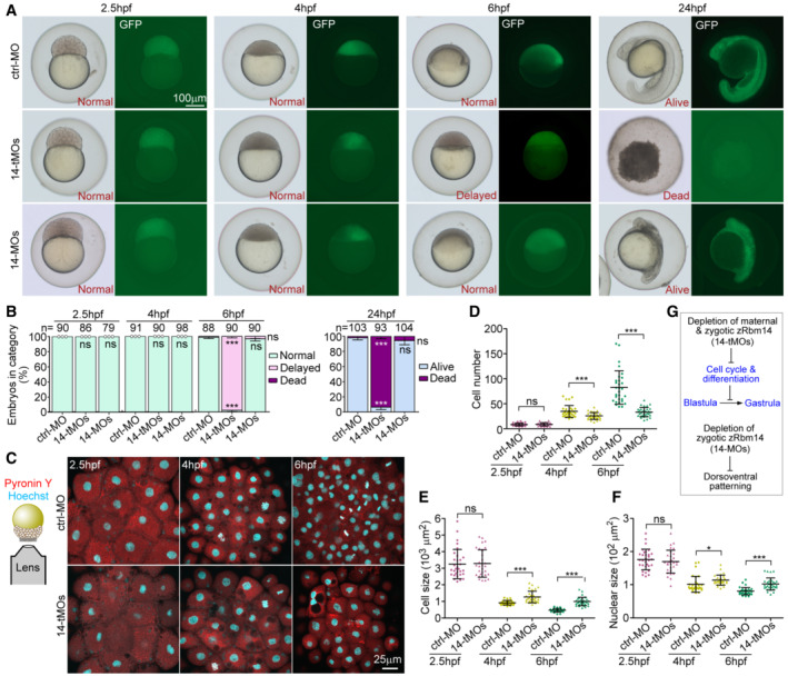
Figure 4. Maternal zRbm14 morphants display impaired cell cycle, blastula arrest, and embryonic death.

-
A, BPhenotypes of zRbm14 morphants. Totally 8 ng of ctrl‐MO, 14‐tMOs, or 14‐MOs were co‐injected with 100 pg of in vitro transcribed GFP mRNA (as a tracer) into 1‐cell embryos to generate control, maternal zRbm14, and zygotic zRbm14 morphants, respectively. Representative embryos, imaged under a dissecting microscope, are shown in (A) and phenotyping results in (B). Total numbers of embryos analyzed are listed over histograms. Sample dots are included for quantification results with no error bars. Please also refer to Fig EV2.
-
C–FMaternal zRbm14 morphants displayed reduced cell proliferation in MZT. Embryos stained with Hoechst 33342 and Pyronin Y to mark the nucleus and the cytoplasm, respectively, were imaged from the animal pole to cover a 10‐μm z‐depth with a confocal microscope (C). Maximum intensity‐projected images (C) from 30 embryos in each condition were used to quantify cell number (D), cell size (E), and nuclear size (F). Each sample dot represents the average value from each embryo.
-
GSummary of the results.
Data information: Quantification results are presented as mean ± SD from three independent experiments. Student's t‐test against ctrl‐MO‐injected populations: ns, no significance (P > 0.05); *P < 0.05; ***P < 0.001.
Source data are available online for this figure.
