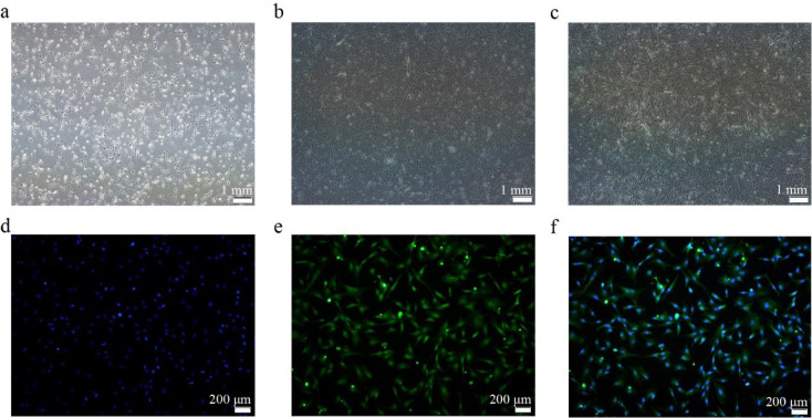
Fig. 1. CS2s separation and identification.
(a) Cell morphology after 24 h of separation. (b) Cell morphology after 36 h of separation. (c) Cell morphology after 48 h of separation. (d) DAPI (blue). (e) PAX7 protein signal (green). (f) Merge image. CS2s, duck myoblasts; DAPI, 4′,6-diamidino-2-phenylindole; PAX7, paired box 7.

