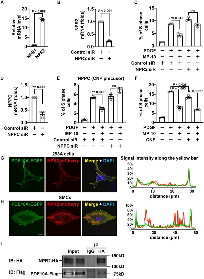
Figure 5.
PDE10A-modulated SMC proliferation is associated with CNP/NPR2 signalling pathway. (A) Relative mRNA levels of NPR1 and NPR2 in rat SMCs. An unpaired Student’s t-test with Welch’s correction was performed. n = 3 for each group. (B) RT-qPCR showing the level of NPR2 in SMCs treated with control or NPR2 siRNA. The expression level was normalized to the control siRNA-treated group. A one-sample t-test was performed to determine the knockdown efficiency. n = 3 for each group. (C) Percentages of S-phase cells treated with 2.5 μM MP-10, NPR2 siRNA, or both. A Welch’s ANOVA with Dunnett’s T3 multiple comparisons test was applied. n = 3 for each group. (D) RT-qPCR results showing the level of NPPC (encoding a precursor of CNP) in SMCs treated with control or NPRC siRNA. The mRNA level was normalized to the control siRNA-treated group. A one-sample t-test was performed to determine the knockdown efficiency. n = 3 for each group. (E) Percentages of S-phase cells treated with 2.5 μM MP-10, NPPC siRNA, or both. A Welch’s ANOVA with Dunnett’s T3 multiple comparisons test was applied. n = 3 for each group. (F) Percentages of rat SMCs in the S-phase after being treated with 2.5 μM MP-10, 1 μM CNP, or both. A Welch’s ANOVA with Dunnett’s T3 multiple comparisons test was performed. n = 3 for each group. (G and H) Confocal microscopic images (left panels) showing the signals of EGFP-tagged PDE10A (green) and mCherry-tagged NPR2 (red) overexpressed in 293A cells (G) or rat SMCs (H). Scale bar = 10μm (G) or 20 μm (H). The intensities of green and red signals across the cell were measured (right panels). (I) Immunoblot showing PDE10A and NPR2 from co-immunoprecipitation. Flag-tagged PDE10A and HA-tagged NPR2 were overexpressed in 293A cells. All data are shown as mean ± SEM. n = 3 for each group. ns, not significant.

