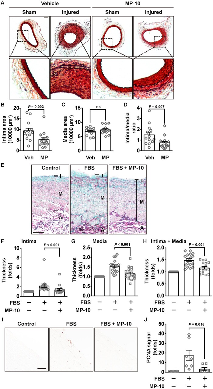
Figure 7.
Neointimal formation in wire-injured mouse femoral arteries and remodelling of HSV segments after vehicle or MP-10 treatment. (A) Representative histological images of cross-sections of sham and injured femoral arteries of control or MP-10-treated mice. Vehicle (40% HP-β-CD) or MP-10 (10 mg/kg/day) was administrated subcutaneously, which started 3 days before the surgery and lasted for 4 weeks. Scale bar = 50 μm. (B) Intimal area, (C) medial area, and (D) the intima/media ratio of mice 4 weeks after the surgery. An unpaired Student’s t-test was used for comparing the media area. Mann–Whitney tests were used for the intima area and the intima/media ratio. n = 13 mice for vehicle treatment and n = 15 mice for MP-10 treatment. (E) Representative images showing HSV wall thicknesses from HSV segments without culture, or with culture in the presence of vehicle or MP-10 (20 μM). HSV segments were cultured in 15% FBS for 2 weeks. Samples were stained by the combined Verhoeff’s elastic and Masson’s trichrome method. Scale bar = 100μm. (F–H) The quantitative data showing thickness of the intimal layer (F), the medial layer (G), or the combined (intima + media) layer (H) of HSV samples from 17 patients after CABG. Thickness from three different locations of each section, and five sections, 500 µm apart, of each sample, were measured and averaged. The total thickness of both the intima and the media layers was calculated and normalized to the uncultured control group. Paired Student’s t-tests were performed. (I) Immunohistochemical staining of PCNA in HSV samples. Scale bar = 50μm. (J) The quantitative data showing the fold change of the PCNA signal in HSV samples. Paired Student’s t-tests were performed. n = 7 (randomly chosen from 17 patient samples). All data are shown as mean ± SEM.

