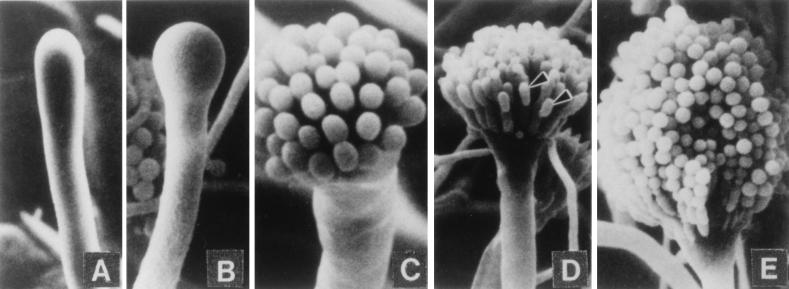
FIG. 1.
Morphological changes during conidiophore formation. Shown are scanning electron micrographs of the stages of conidiation. (A) Early conidiophore stalk. (B) Vesicle formation from the tip of the stalk. (C) Developing metulae. (D) Developing phialides. (E) Mature conidiophores bearing chains of conidia. Reproduced from reference 109 with permission of the publisher.

