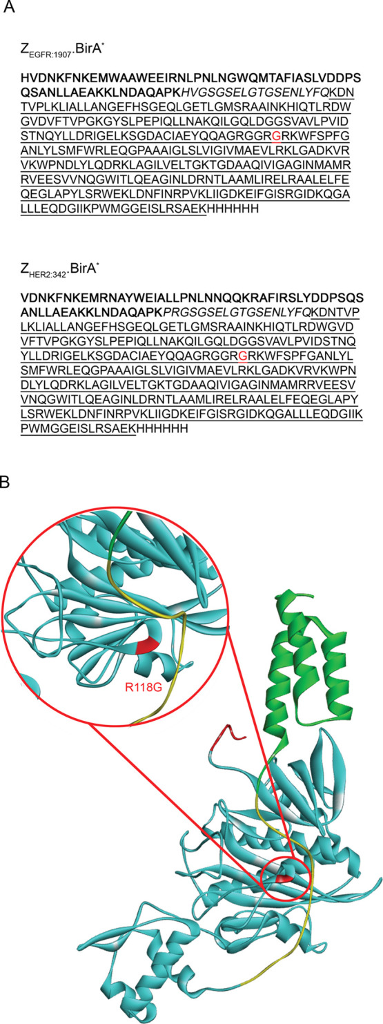
Figure 1.

Design of the Affibody.BirA* fusion constructs, ZEGFR:1907.BirA* and ZHER2:342.BirA*. (A) Amino-acid sequences of ZEGFR:1907.BirA* (top) or ZHER2:342.BirA* (bottom). The affibody ligand sequence (in bold characters) is followed by a linker region (italics), a C-terminal BirA* catalytic domain (underlined), and ending with a hexahistidine tag. (B) Ribbon representation of the Affibody.BirA* fusion construct, ZEGFR:1907.BirA* generated from the I-TASSER server.17 Color pattern: affibody ligand (in green), linker containing a TEV cleavage site (in yellow), C-terminal BirA* catalytic domain (in cyan), and a histidine tag (in red). The location of the R118G mutation in BirA* is highlighted in red (magnified image).
