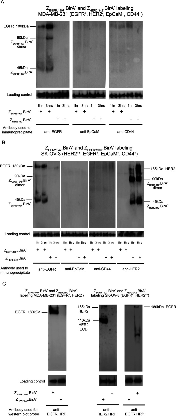
Figure 4.

ZEGFR:1907.BirA* and ZHER2:34.BirA* are able to biotinylate EGFR and HER2, respectively, on the surface of human cancer cells expressing EGFR or HER2. (A) EGFR-expressing cells (MDA-MB-231) were only biotinylated with ZEGFR:1907.BirA* or ZHER2:342.BirA*. Labeled cells, at 1 h and 3 h time points, were lysed, and EGFR itself was pulled down (IP) using Cetuximab (human EGFR mAb). The captured biotinylated EGFR was detected by Western blot using streptavidin-HRP. (B) The same experiment was performed for ZHER2:342.BirA* and ZEGFR:1907.BirA* using the HER2-overexpressing and EGFR-expressing cells (SK-OV-3). Specifically, cells were biotinylated with either ZHER2:342.BirA* or ZEGFR:1907.BirA*. Cells were collected at 1 h and 3 h time points, lysed, and HER2 and EGFR were pulled down (IP) using Trastuzamab (human HER2 mAb) or Cetuximab (human EGFR mAb), respectively. The captured biotinylated HER2 or EGFR were detected by Western blot using streptavidin-HRP. For both cell lines, MDA-MB-231 (A) and SK-OV-3 (B), EpCaM, and CD44 were also pulled down, and their lack of biotinylation was confirmed in order to establish the specificity of the biotinylation event. For panels A and B, immunoprecipitation elution samples served as loading controls, probed with mAbs specific for EGFR, HER2, EpCaM, and CD44 to confirm that an equal amount of each marker was loaded on the beads. (C) MDA-MB-231 and SK-OV-3 cells were biotinylated with either ZEGFR:1907.BirA* or ZHER2:342.BirA*, as previously described for 3 h. Cells were then lysed, and biotinylated protein species were pulled down using streptavidin dynabeads. The captured biotinylated proteins were analyzed by Western blot. In the case of the EGFR-expressing MDA-MB-231 and SK-OV-3 cells, biotinylated EGFR was detected using an anti-EGFR-HRP conjugate. An anti-HER2.HRP conjugate was used to detect biotinylated HER2 recovered from HER2-expressing SK-OV-3 cells. Western blots of cell lysates probed with either anti-HER2 or anti-EGFR mAbs served as the loading controls of these markers. SDS-PAGE and Western blots were performed on samples prepared under nonreducing conditions, resulting in the observation of Affibody.BirA* monomers and dimers.
