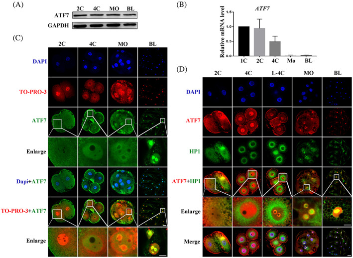
FIGURE 1.

Expression and localization of ATF7 during porcine embryonic development. (A) Western blotting results of ATF7 protein expression levels during early porcine embryonic development. (B) Real‐time quantitative PCR results of ATF7 mRNA expression levels during early porcine embryonic development. (C)ATF7 antibody to detect ATF7 localization during early porcine embryonic development. At the 2C stage, ATF7 was enriched near the nucleus. At the 4C stage, ATF7 entered the nucleus. When embryos were at the MO and BL stages, ATF7 was mainly localized on pericentric heterochromatin. Blue, DAPI; red, TO‐PRO‐3; green, ATF7; bar = 20 μm. (D) Typical picture of localization of ATF7 and HP1. Colocalization of ATF7 and HP1 was evident from the late 4‐cell to the BL stage. Blue, DAPI; red, ATF7; green, HP1; bar = 20 μm
