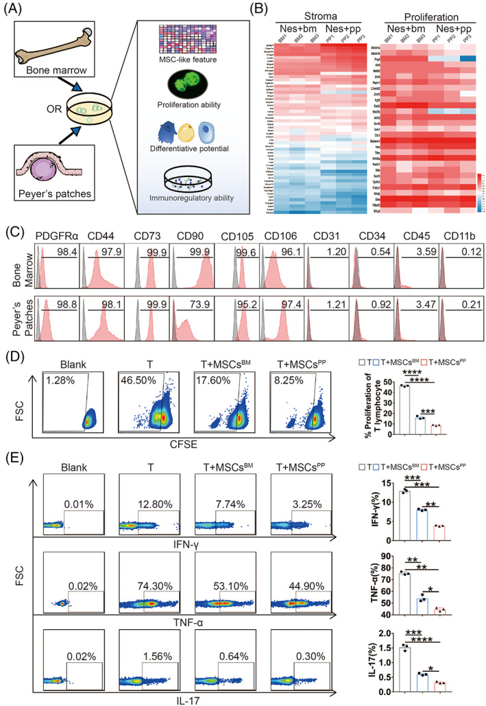
FIGURE 1.

Isolation and characterization of Nestin+ cells derived from bone marrow or Peyer's patches in transgenic mice. (A) A graphical scheme of Nestin+ cell isolation. (B) Heat map showing the expression patterns of representative genes with relevant functions. (C) Expression of MSC‐related markers on bone marrow‐ and Peyer's patch‐derived Nestin+ cells. The proliferation (D) and secretion of IFN‐γ, TNF‐α, and IL‐17 (E) from splenic CD3+ T cells were analysed after T cells were cocultured with or without MSCsBM/PP. Data were presented as the means ± SD (n = 3). *p < 0.05, **p < 0.01, ***p < 0.001, ****p < 0.0001. TNF, tumour necrosis factor; IFN, interferon; IL, interleukin; MSCsBM, bone marrow‐derived Nes + MSCs; MSCsPP, Peyer's patch‐derived Nes + MSCs.
