. 2023 Jan 11;5(3):571–595. doi: 10.1039/d2na00483f

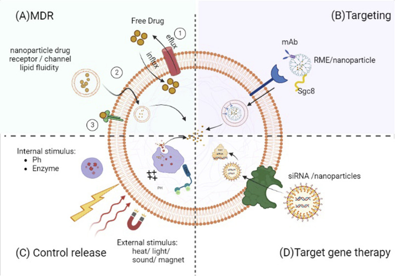
Fig. 2. Control release: drug content liberation from nanocarriers can be stimulated by certain microenvironmental parameters at the target location (e.g., pH change and enzymatic activities) or by external stimuli (e.g., heat, light, electric and magnetic field or ultrasound). Targeting: targeting ligands can specifically attach to the malignant cell (e.g., monoclonal antibodies (mAbs) or sgc8 aptamer). Targeted gene therapy: nanoparticle formation of targeted gene therapy agents (e.g., siRNA and TKIS) increased their anti-leukemic potency by enhancement of their stability and/or concentration. MDR (multi-drug resistance). Free small drugs move into malignant cells through passive transmission across the cell membrane, and they come into contact with membrane proteins such as drug efflux pumps leading to less than optimum amounts in the target cell. Lipid packing density and velocity can affect diffusion through the membrane. Increased membrane fluidity implies considerable drug permeability by delivering therapeutic agents into cancer cells without relying on specific receptors or channels, and nanoparticles can circumvent this resistance mechanism.

Fig. 2