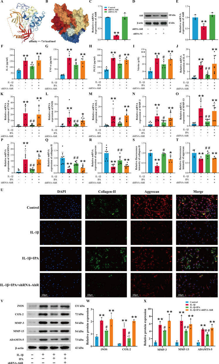
Fig. 4.
Protective effects of IPA depend on AhR. A The binding mode and secondary structure of AhR were demonstrated by a cartoon model. B The binding of the AhR pocket was shown with a space-filling model. C RT-qPCR analysis of AhR (n = 3); D, E Western blot and semi-quantitative analysis of AhR (n = 3). F–H Expression of IL-6, TNF-α and PGE2 was detected by ELISA (n = 5). I Expression of NO was detected by Griess reaction (n = 5). J–R RT-qPCR analysis of IL-6, TNF-α, iNOS, COX-2, MMP-3, MMP-13, ADAMTS-5, collagen-II and aggrecan (n = 9); S–U Immunofluorescence and semi-quantitative analysis of aggrecan and collagen-II (n = 5, scale bar = 100 μm, ×200); V–X Western blot and semi-quantitative analysis of COX-2, iNOS, MMP-3, MMP-13 and ADAMTS-5 (n = 3). *p < 0.05, **p < 0.01 vs. the control group, #p < 0.05, ##p < 0.01 vs. the IL-1β group

