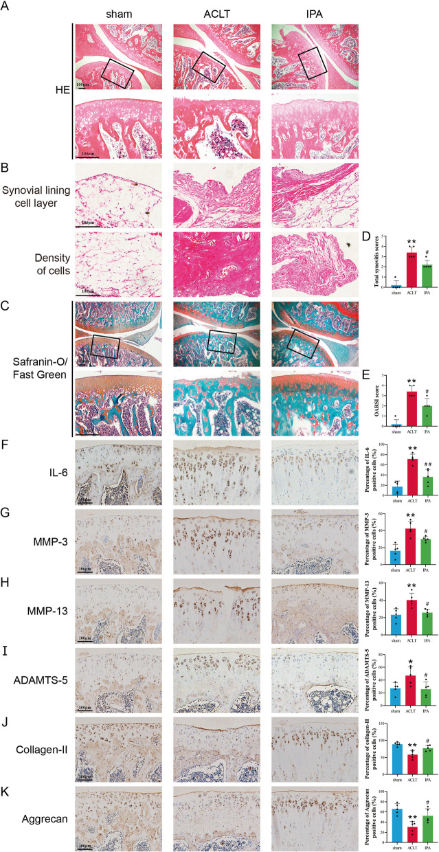
Fig. 6.
IPA attenuated OA progression in rat model. A–C Representative images of safranin O-fast green and H&E staining of cartilage and synovial sections from different groups. D The total synovitis scores of different groups. E The cartilage OARSI scores of different groups. F–K Immunohistochemistry staining and percentage of positive cells of MMP-3, MMP-13, ADAMTS-5, aggrecan and collagen-II. n = 5, Scale bar = 100 μm. **p < 0.01 vs. the control group, ##p < 0.01 vs. the ACLT group

