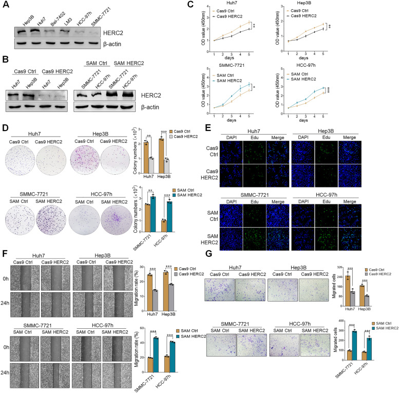
Fig. 2.
HERC2 promoted the malignant phenotype of HCC cells. A Expression levels of HERC2 in Hep3B, Huh7, Bel-7402, LM3, HCC-97 h, and SMMC-7721 cell lines were measured by immunoblotting assay. B Western blot analysis of HERC2 protein levels both in HERC2 knockout and overexpression cell lines. C A CCK-8 assay was used to detect cell proliferation in both HERC2 knockout and HERC2 overexpression cell lines. D A colony formation assay was performed to investigate cell proliferation in HERC2 knockout and overexpression cell lines. E Immunofluorescent detection of EdU was employed to determine the proliferation of HERC2 knockout and overexpression cell lines. Wound healing assays F and migration tests G were used to detect the migration ability of HERC2 knockout and overexpression HCC cells. *p < 0.05, **p < 0.01, ***p < 0.001. Data from one representative experiment of three independent experiments are presented

