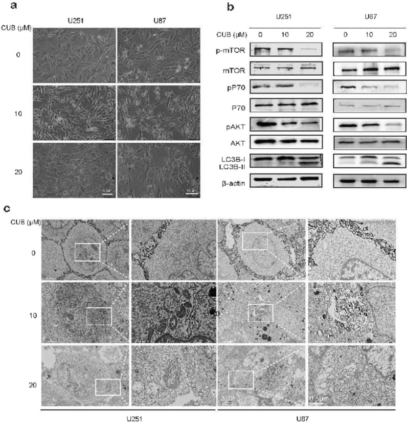
Fig. 3.
CUB induces GBM cell autophagy. a Morphological images of GBM cells. b Western blot analysis detected markers of the Akt/mTOR/pS70k pathway and LC3B-I, LC3-II in U251 cells treated with CUB at indicated concentration for 24 h (10 µg of the cell lysates was loaded)(Cropped blots, original cropped images and replicates are presented in Additional file 1: Fig. S5 and S6). c SEM images of U251, U87 cells treated with CUB (10, 20 µM) or DMSO for 24 h. Boxes highlight autophagic vacuoles and ER stress bubbles. Scale bars were marked in the images

