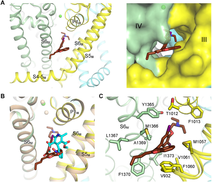
Figure 2 ∣. Coordination of AMIO in rCav1.1.
(A) AMIO inserts into the fenestration on the interface of repeats III and IV (the III-IV fenestration). The PD of rCav1.1A is shown as ribbon cartoon (left) or semi-transparent surface (right) to highlight the binding pocket for AMIO. (B) AMIO occupies the same binding site as the DHP compounds. Structures of the α1 subunit of rCav1.1A and nifedipine (cyan sticks)-bound rCav1.1 (colored wheat, PDB code: 6JP5) can be superimposed with RMSD of 0.57 Å over 970 aligned Cα atoms. (C) AMIO is coordinated exclusively through van der Waals contacts. AMIO and the surrounding residues are shown as sticks. See also Figure S2.

