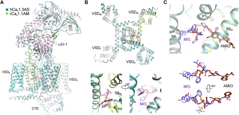
Figure 4 ∣. AMIO and SOF interact within human Cav1.3 (hCav1.3).
(A) The overall structure of hCav1.3 bound to AMIO and SOF (hCav1.3AS) is identical to that of rCav1.1AM. The two structures can be superimposed with an RMSD of 0.66 Å for 1782 Cα atoms in the α1 and α2δ-1 subunits. rCav1.1AM is domain colored and hCav1.3AS is colored dark green. (B) SOF and MNI-1 share identical binding pose in the presence of AMIO. Top: A cytosolic view of superimposed α1 subunits from hCav1.3AS and rCav1.1AM. Bottom: Two opposite side views of superimposed α1 subunits from the two structures. (C) Direct interaction of SOF and AMIO within hCav1.3AS. SOF (pink) and MNI-1 (light purple), with similar chemical structures, interact with AMIO in the same way within the PD of LTCCs. See also Figures S4, S5.

