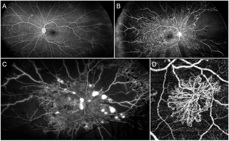
Figure 1.
Angiographic findings in different stages of diabetic retinopathy. (A) Mild form of NPDR characterised by preserved posterior pole and minor capillary non-perfusion changes detected in the extreme periphery. (B) Advanced form of NPDR, showing extensive peripheral capillary non-perfusion and central involvement with several microaneurysms and macular edoema. (C) A case of PDR with extensive peripheral capillary non-perfusion and neovascularizations detected both at the level of the optic nerve head and the retinal periphery. (D) Optical coherence tomography angiography reconstruction of a PDR-related neovascularization.

