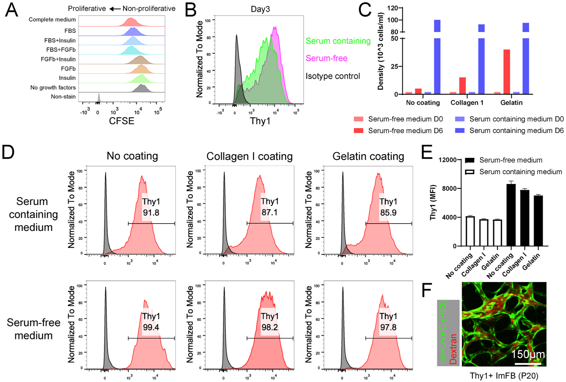
Fig. 7.

FBs maintain Thy1 expression in serum-free culture medium. (A) Representative histograms showing the proliferation of ImFBs cultured in the FB medium (Fibrolife) supplemented with different growth factors. ImFBs were stained with CFSE as proliferation indicator. (B) Representative histograms showing the Thy1 expression in ImFBs cultured in serum-containing medium or serum-free medium for 3 days. (C) Statistical quantification of Thy1+ ImFB cell density cultured in serum-containing medium or serum-free medium on day 0 (D0) and day 6 (D6). (D, E) Representative histograms (D) and statistical analysis (E) showing the Thy1 expression in ImFBs cultured in serum-containing medium or serum-free medium on collagen I- or gelatin-coated surfaces. (F) Representative images of μVNs made of ImHUVECs (P23, green) with Thy1+ ImFB (P20). μVNs are perfused with Texas Red dextran (red). Scale bar is 150 μm.
