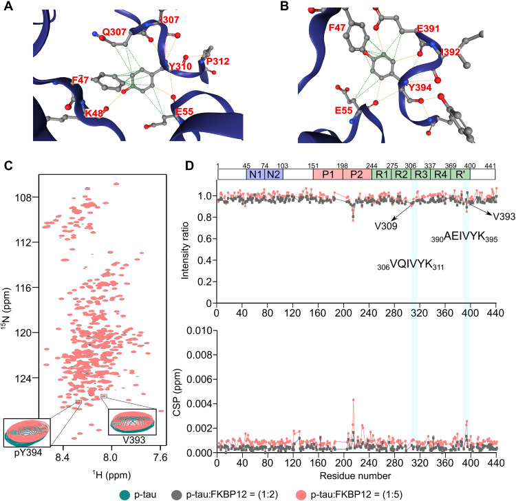
Fig. 6. Tyrosine phosphorylation blocks tau interaction with FKBP12.
(A and B) Contacts between Y310 (A) or Y394 (B) of tau with F47 and E55 of FKBP12. (C) 2D 1H-15N HSQC spectra of cAbl-phosphorylated tau in the absence (green) or fivefold excess (brick red) of FKBP12. The NMR signals of V393 and pY394 are highlighted in the inset. (D) Changes in the intensities (top) and CSPs (bottom) of cross peaks in the HSQC spectra of cAbl-phosphorylated tau upon addition of twofold (gray) or fivefold (brick red) excess of FKBP12. The domain organization of tau is shown on top.

