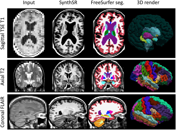
Fig. 1. Examples of inputs and outputs of SynthSR from the MGH dataset.
Each of the inputs has been acquired with a different orientation (axial, sagittal, and coronal), slice spacing (6, 5, and 4 mm), and MR contrast (turbo spin echo T1, T2, FLAIR); we visualize them in orthogonal view to illustrate their low-resolution out of plane. SynthSR produces a synthetic 1-mm isotropic MPRAGE in all cases, which is compatible with nearly every existing brain MR image analysis method. The third column shows the automated segmentation of the synthetic MPRAGE obtained with FreeSurfer, and the last column shows three-dimensional (3D) renderings of (top to bottom) the subcortical segmentation, parcellated white matter surface, and parcellated pial surface. We emphasize that all images were processed with the same neural network using SynthSR out of the box, without retraining.

