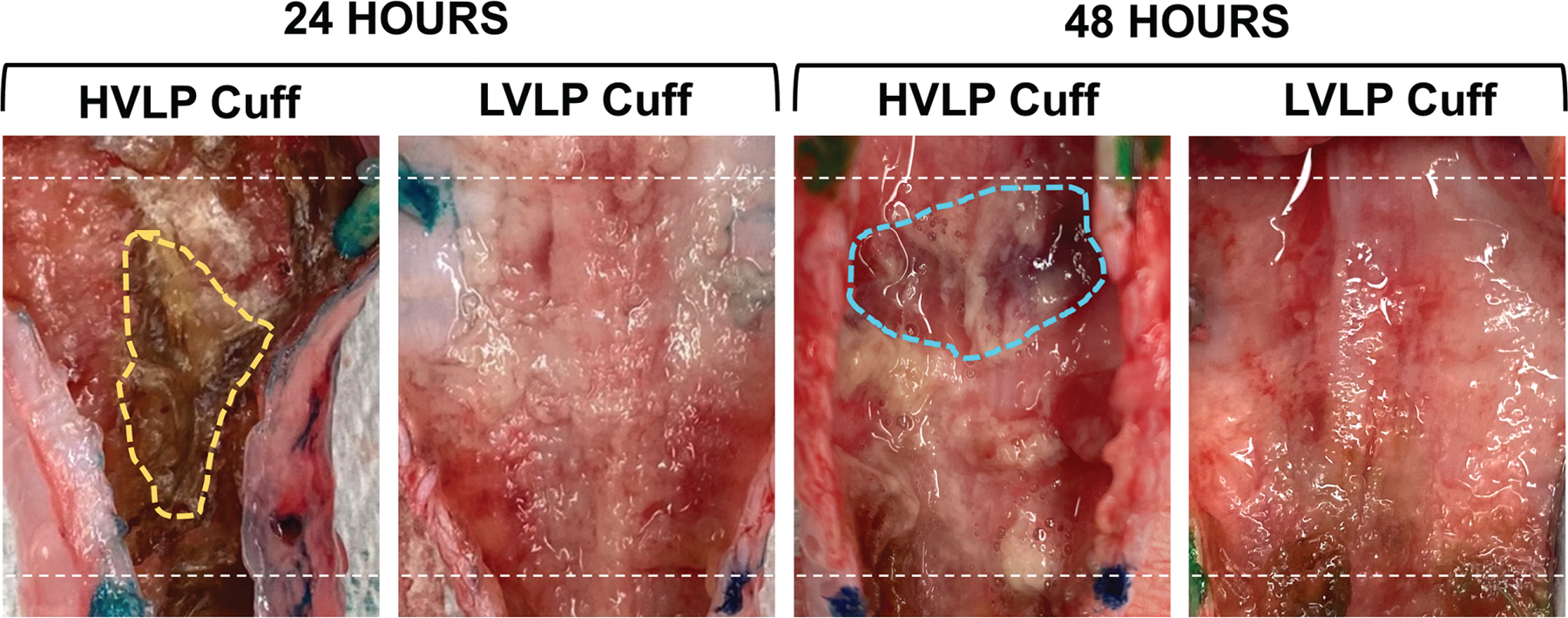
Figure 2.

Gross tracheal specimen mucosa following tracheostomy cuff placement. Following 24 and 48 hours, there was marked necrotic debris (yellow) and edematous tissue (blue) noted in high-volume, low-pressure trachea mucosa. Dotted line indicates proximal/distal cuff margins. HVLP, high volume, low pressure; LVLP, low volume, low pressure.
