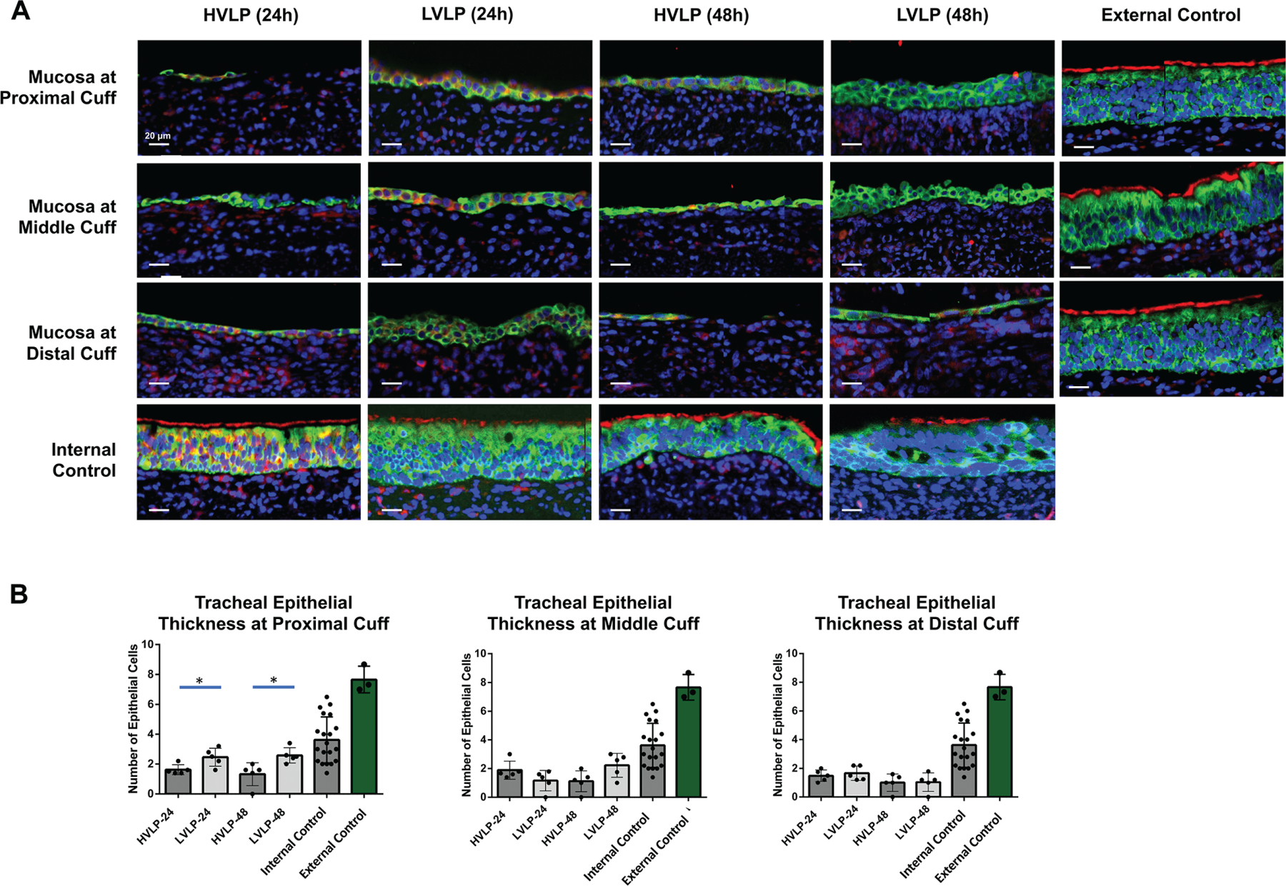
Figure 3.

Tracheal mucosa at cuff contact compared to control. (A) Representative TUBA/panCK staining in normal pig trachea with uninjured ciliated epithelium vs cuff. (B) Epithelial cell thickness following 24-hour/48-hour cuff contact compared to controls. *P <. 05. HVLP, high volume, low pressure; LVLP, low volume, low pressure.
