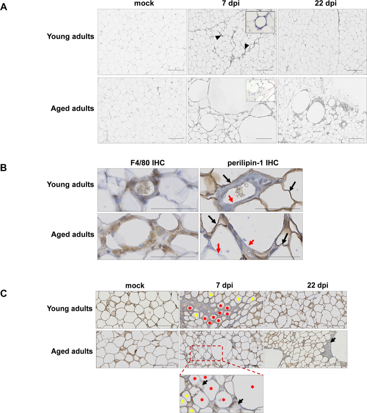
Fig. 4. SARS-CoV-2 infection induces adipocyte death in the SCAT, and dead adipocytes are not cleared in aged golden hamsters.
A Representative microscopy images of the (inguinal) SCAT of young adult and aged hamsters at 0 (mock), 7, and 22 dpi (H&E staining). At 7 dpi, one can note the presence of CLSs in the SCAT of young-adult animals, and the presence of fibrosis in the SCAT of the aged animals (inserts: higher magnification). B Representative immunochemical (IHC) staining for F4/80 (left panels) highlighted the presence of macrophages (in brown) clustered around adipocytes. Representative IHC staining for lipid-droplet-specific perilipin-1 (right panels) highlighted live (stained) adipocytes (in brown, black arrows) and damaged/dead (non-stained, red arrows) adipocytes in the SCAT of young adults and aged adults at 7 dpi. C Representative images of dead adipocytes in the SCAT of young adult and aged hamsters at 0 (mock), 7, and 22 dpi. IHC staining for perilipin-1 shows live (stained) adipocytes. A higher magnification image is shown for aged adult animals at 7 dpi. Live adipocytes are indicated by yellow stars, and damaged/dead adipocytes are indicated by red stars. Lipid spillovers are indicated by black arrows. A–C scale bars = 100 μm. B, C Slides were counterstained with Mayer’s hematoxylin.

