
Figure 6.
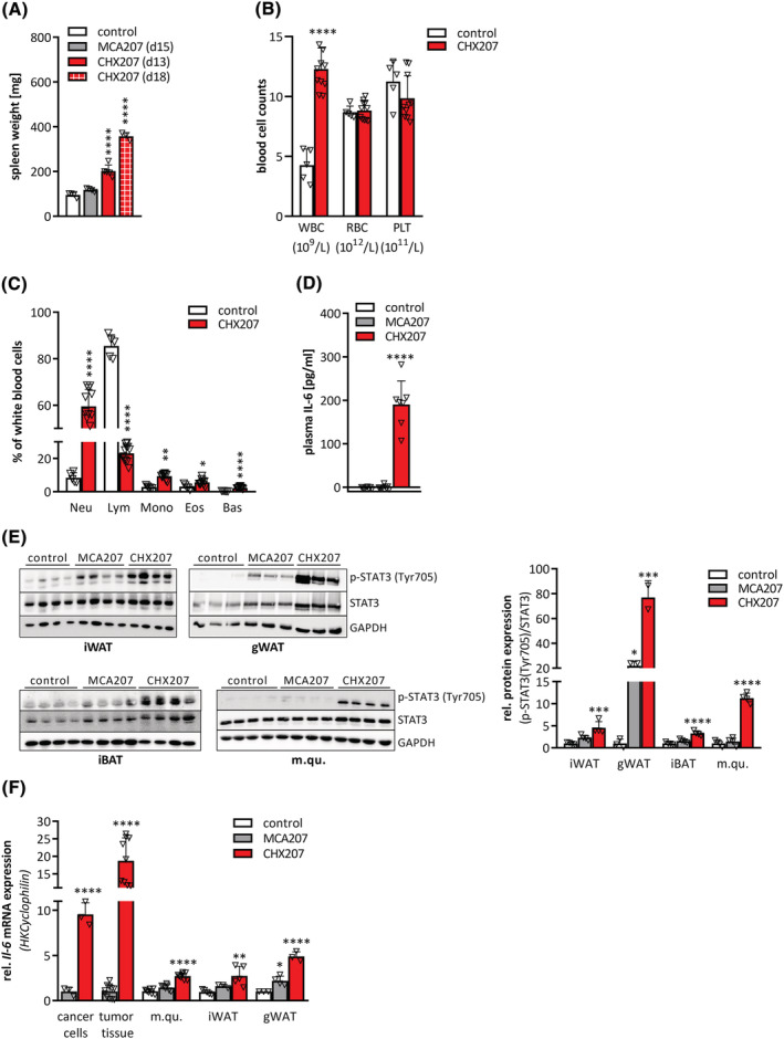
CHX207 mice exhibit systemic inflammation and high levels of circulating IL‐6. (A–F) Ten‐ to 11‐week‐old male C57BL/6J mice were injected with 1 × 10⁶ MCA207 cells, 1 × 106 CHX207 cells, or 1× PBS (control). (A) Mice were sacrificed at the indicated timepoints, spleens were excised and weighed. (B,C) Cell counts of whole blood from control and tumour‐bearing mice were analysed using an abacus Haematology analyser (n = 5–11, Day 14 p.i.). (B) Absolute counts of white blood cells (WBC), red blood cells (RBC), and platelets (PLT). (C) Relative white blood cells subtypes (neutrophils (Neu), lymphocytes (Lym), monocytes (Mono), eosinophils (Eos), basophils (bas)). (D) Plasma IL‐6 concentrations were determined using ELISA (n = 7–9). (E) Western blotting analysis of p‐STAT3 (Tyr705) and STAT3 protein expression and quantification of p‐STAT3 (Tyr705) relative to total STAT3 in inguinal white adipose tissue (iWAT), gonadal white adipose tissue (gWAT), interscapular brown adipose tissue (iBAT), and musculus quadriceps (m.qu.) (Day 13 p.i.). GAPDH was used as loading control. (F) mRNA expression levels of Il‐6 in MCA207‐ and CHX207‐cancer cells (n = 3–4) and tumour tissue, m.qu., iWAT and gWAT (Day 13 p.i., n = 7–11) were determined by qRT‐PCR. Cyclophilin was used as housekeeping gene. Data are presented as means + SD. Significance was determined by B,C,F) two‐sided Student's t‐test or A,D,E,F) one‐way ANOVA (*P ≤ 0.05, **P ≤ 0.01, ***P ≤ 0.001, ****P ≤ 0.0001).