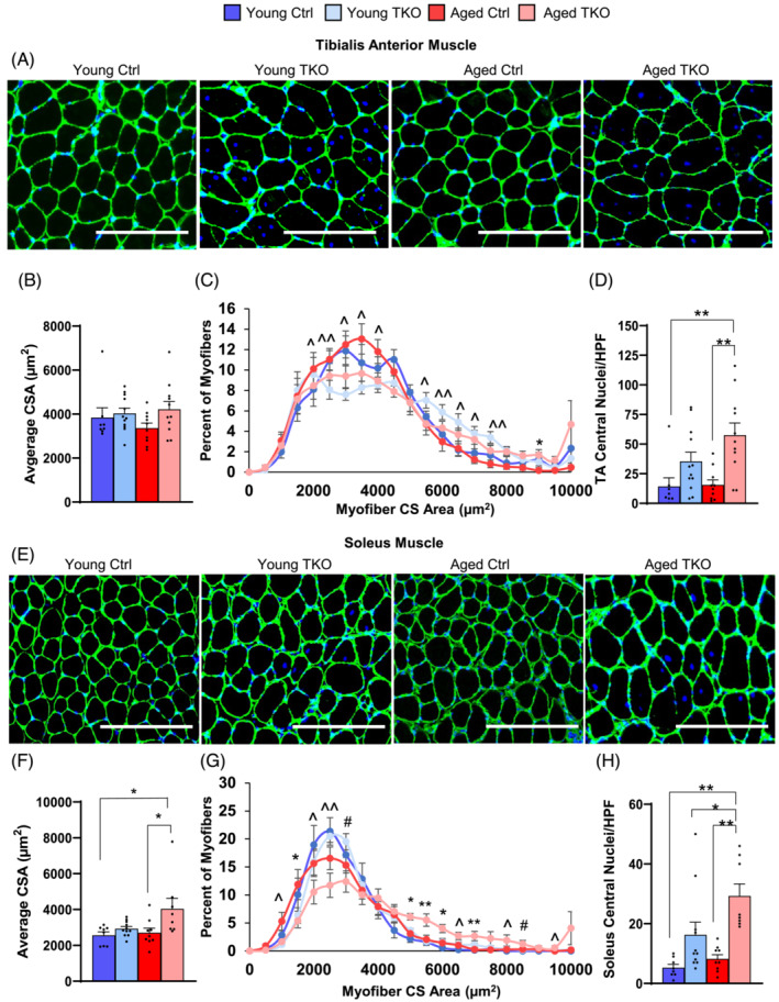
Figure 2.

FoxO deletion in muscle mildly increases fibre size and markedly increases central nuclei in TKO muscle. Laminin and DAPI stain of tibialis anterior (TA) cross sections (scale bar 250 μm) (n = 8–13 per group) (A). Average cross sectional area (CSA) (B) and distribution of CSA (C) from TA. Quantification of fibres with central nuclei of TA muscle are shown (D). Laminin and DAPI stain of soleus cross sections (scale bar 250 μm) (n = 8–13 per group) (E). Average CSA (F) and distribution of myofibre CSA (G) from soleus. Quantification of central nucleated fibres in soleus (H). HPF, high power field. *P < 0.05; **P < 0.01 Aged Ctrl vs. Aged TKO or as indicated; ^P < 0.05, ^^P < 0.01 genotype main effect and #P < 0.05 age main effect by 2‐way ANOVA.
