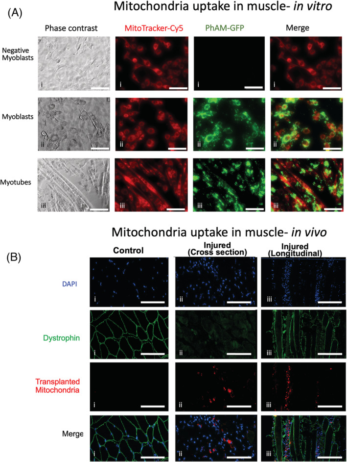
Figure 2.

Uptake of donor mitochondria in myoblasts, myotubes and myofibres. (A) MitoTracker deep red was incubated on C2C12 cells to label resident mitochondria red (Cy5). Donor PhAM (green) mitochondria were isolated from Dendra‐2 mice and incubated with MitoTracker‐labelled C2C12 cells (i,ii) or myotubes (iii) at 37°C. C2C12 cells were grown to 70% confluency then differentiated into myotubes for 5 days using 2% horse serum (iii). Negative myoblasts (i) did not receive donor mitochondria. The cells were imaged 24 h later. Green donor mitochondria can be seen incorporated into the host C2C12 myoblasts and myotubes. Many donor mitochondria appear to be perinuclear. The scale bar corresponds to 200 μm. (B) Examples of mouse gastrocnemius tissue sections from mitochondrial transplant therapy (MTT) mice that were undamaged control muscles injected with phosphate‐buffered saline (PBS) (i) or 2 days following injection with BaCl2 to induce muscle injury (ii,iii). Examples of injured muscles are shown in cross section (ii) or longitudinal section (iii). Mitochondria from C57BL/6 mice were isolated and labelled with MitoTracker deep red then injected into the tail vein of mice, 24 h after the BaCl2 injury in one limb. Contralateral control (undamaged gastrocnemius muscles) in MTT mice (i) and BaCl2 injured muscle was cut at a thickness of 8 μm in cross section (ii) or longitudinal section (iii) and reacted with antibodies to dystrophin to identify the muscle sarcolemma and counter stained with DAPI to identify myonuclei. MitoTracker deep red stained the donor mitochondria red and where imaged in the Cy5 channel. The data show no red‐stained mitochondria that were taken up by the contralateral control muscle (i), but some fibres that were damaged (as shown by disruption of the dystrophin in the fibre sarcolemma) took up the systemically delivered mitochondria. Longitudinal sections (iii) showed evidence of damaged fibres that has taken up labelled donor mitochondria. The scale bar corresponds to 200 μm.
