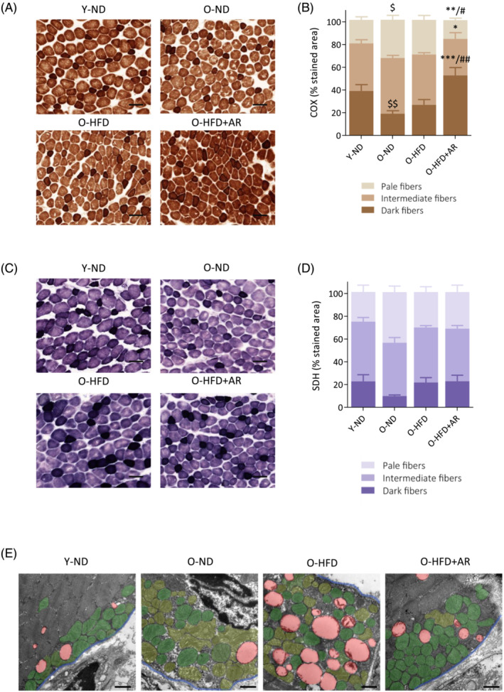
Figure 7.

Chronic administration of AdipoRon enhances mitochondrial function and protection in muscle of middle‐aged obese mice. (A,C) Histochemistry staining of COX and SDH activity in the rectus femoris of the four groups mice, the darkest colour being associated with the highest activity. Representative images of both stainings for each group are shown. Scale bar = 100 μm. (B,D) quantification of COX and SDH activity. Activity was expressed as the percentage of stained area normalized to the cross‐sectional area of the muscle, for each of the three staining intensities (set up as corresponding to pale, intermediate or dark colour). Data are means ± SEM for 4–6 mice per group. Statistical analysis was performed by one‐way ANOVA (comparing three groups of O‐mice) or by unpaired two‐tailed t‐test (Y‐ND vs. O‐ND). $ P < 0.05, $$ P < 0.01 versus Y‐ND mice. *P < 0.05, **P < 0.01, ***P < 0.001 versus O‐ND mice. # P < 0.05, ## P < 0.01 versus O‐HFD mice. (E) Transmission electron micrographs of mitochondria in the subsarcolemmal region of rectus femoris in the four groups of mice. For the sake of clarity, abnormal mitochondria are false‐coloured in pale green, while normal mitochondria are coloured in dark green, LDs in red and the blue line delimits the sarcolemma. Representative images of each group are shown. Scale bar = 1 μm.
