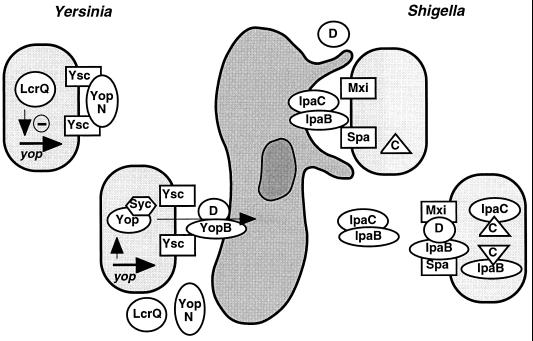
FIG. 8.
Schematic representation of regulation of type III secretion by contact with a eukaryotic cell (shown in the middle) in Yersinia and Shigella spp. For Yersinia spp., prior to cell contact (upper left), the type III secretion channels (Ysc) are kept shut by YopN, and cytoplasmic accumulation of the negative regulatory factor LcrQ leads to transcriptional repression of yop genes (see the text). After cell contact, YopN is released, allowing rapid secretion of LcrQ, which in turn relieves the block from yop gene expression. Anti-host Yop proteins, protected by cognate chaperones (Syc), are translocated into the eukaryotic cell via the type III secretion machinery and YopB/D. For Shigella spp. (only the model of regulation via IpaB/D is shown [see the text for further details]), before contact with a eukaryotic cell (lower right), the S. flexneri type III secretion channels (Mxi and Spa) are kept shut by a complex of IpaB and IpaD. Ipa proteins accumulate in the cytoplasm and are protected from interaction and proteolytic degradation by the chaperone IpgC (C in a triangle). IpaB and IpaC, however, are partially secreted and form a protein-protein complex in the bacterial supernatant. After target cell contact, the IpaB/D block is released and high local concentrations of the IpaB/C complex induce membrane ruffling and uptake of Shigella spp.

