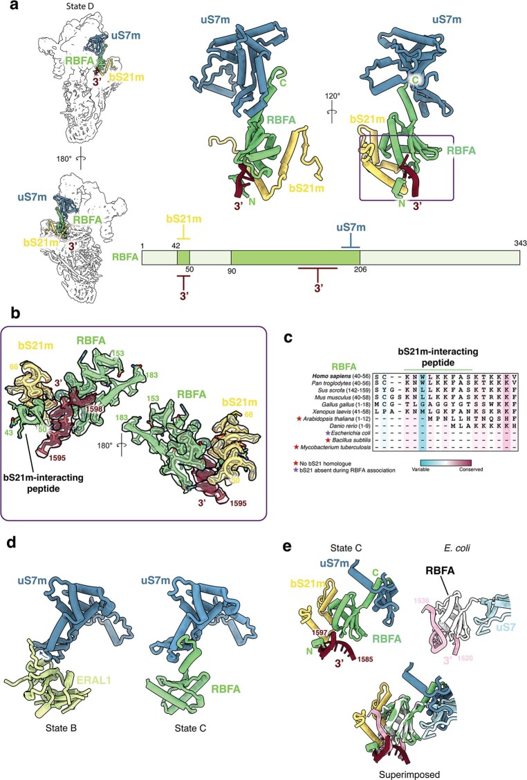
Extended Data Fig. 6. The 3′ rRNA binding mode of RBFA is distinct from that of E. coli.
(a) Interaction network of RBFA in State C in context with the entire complex (left), shown in cartoon (right) and in a schematic (bottom). (b) Model and cryo-EM map of the highlighted region in (a), showing the composite binding site formed by RBFA and bS21m. (c) Conservation analysis of the N-terminal peptide segment observed to interact with bS21m during 3′ rRNA binding. (d) Model illustrating conformational changes in uS7m during RBFA/ERAL1 exchange in States B and C. (e) Comparison of the 3′ rRNA binding mode between human and bacterial systems (PDB:7BOH)23.

