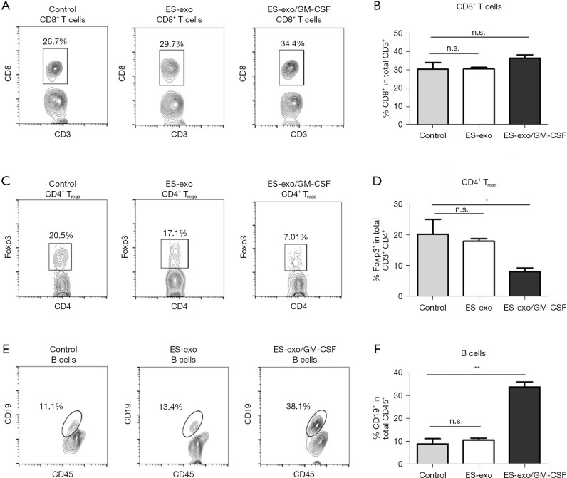
Figure 4.
Vaccination of ES-exo/GM-CSF inhibits tumor-infiltrating Tregs and increases tumor-infiltrating B cells in metastatic lung tumors. Female C57BL/6 mice were vaccinated twice (days 0 and 7) with PBS (control) or ES-exo or ES-exo/GM-CSF prior to tail vein injection with LLC cells on day 14. Five weeks following LLC challenge, lung tumors were resected, digested by enzymes, and tumor-infiltrating cells were harvested and analyzed by flow cytometry. The pan-hematopoietic marker CD45 was used to identify intratumoral immune cells. (A) The presence of tumor-infiltrating cytotoxic T cells (Tc) was examined and the percentages of CD8+ Tc cells in CD3+ T cell population were determined. (B) Bar graphs showing percentages of tumor-infiltrating CD8+ Tc cells in CD3+ T cells (n=4 control and ES-exo group, and n=6 mice in ES-exo/GM-CSF group). Mean ± SD, ANOVA with Tukey’s multiple comparison test. (C) Tumor-infiltrating Foxp3+ Tregs in CD3+ CD4+ T cells obtained from control, ES-exo and ES-exo/GM-CSF-vaccinated mice were evaluated. Numbers in the plots represent the percentages of subpopulations. (D) Summary of the data shown in (C). n=4 in control and ES-exo group, n=6 in ES-exo/GM-CSF group; mean ± SD, *, P<0.05; ANOVA with Tukey’s multiple comparison test. (E) Dot plots showing the percentages of CD19+ B cells in CD45+ intratumoral cells obtained from control, ES-exo and ES-exo/GM-CSF-vaccinated mice. Numbers in the dot plots are the percentages of each subpopulation. (F) Bar graphs showing average of percentages of CD19+ B cells in tumor-infiltrating CD45+ cells acquired from control, ES-exo and ES-exo/GM-CSF-vaccinated mice (n=4 mice in control and ES-exo group, n=6 mice in ES-exo/GM-CSF group). Mean ± SD, **, P<0.01; ANOVA with Tukey’s multiple comparison test. ES, embryonic stem; GM-CSF, granulocyte-macrophage colony stimulating factor; n.s. not significant; PBS, phosphate buffered saline; LLC, Lewis lung carcinoma; SD, standard deviation; ANOVA, analysis of variance.

