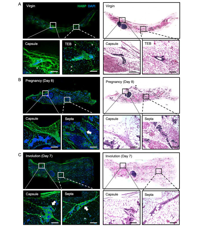
Fig. 1.
Organized hyaluronan-rich septa are deposited throughout the developing mammary gland. Immunofluorescence microscopy for hyaluronic acid binding protein (HABP; green) and DAPI nuclear stain alongside serial hematoxylin and eosin- stained images identifying key hyaluronan (HA) structures within mammary glands of virgin/nulliparous (A), pregnant (B), and involuting (C) BALB/c mice. HA extends throughout the mammary fat pad (IF images), dividing the gland into distinct lobules and encasing it in a fibrous capsule (inserts). Arrows highlight epithelial buds enveloped by HA-rich septa. Asterisks highlight adipocytes with a prominent pericellular HA coat. Whole-gland images were acquired on Leica DM6000B (IF) and DM5500B (H&E) microscopes at 100× magnification and stitched together via the LAS V3.8 software. Inserts were acquired on Leica DM6000B (IF) and DM5500B (H&E) microscopes at 200× magnification. Scale bars represent 100 µM

