Fig. 4.
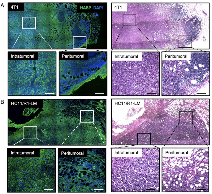
Hyaluronan is heterogeneously deposited throughout mammary tumors. Immunofluorescence microscopy for hyaluronic acid binding protein (HABP; green) and DAPI nuclear stain alongside hematoxylin and eosin (H&E) - stained images identifying hyaluronan (HA) deposition within three murine models of breast cancer: (A) 4T1 and (B) HC11/R1-LM. H&E images of 4T1 and HC11/R1-LM tumors highlight the highly cellular tumor adjacent to the normal, adipocyte-rich mammary gland. Inserts identify intratumoral and peritumoral regions within each tumor. Whole tumor images were acquired on Leica DM6000B (IF) and DM5500B (H&E) microscopes at 200× and 100× magnification, respectively, and stitched together via the LAS V3.8 software. Inserts were acquired on Leica DM6000B (IF) and DM5500B (H&E) microscopes at 20× magnification. Scale bars represent 100 µM

