
FIGURE 1.
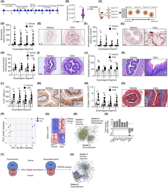
Experimental EoE is characterized by predominant type 2‐associated inflammation that overlaps with the human disease. Schematic representation of the experimental EoE protocol is shown (A). Allergen sensitization was confirmed by quantitation of serum IgE (B). Esophageal specimens (C) were stained with anti‐major basic protein (MBP, E,G) and total (D) as well as intraepithelial (F) eosinophils were quantified. The slides were stained with H&E (I,K), and lamina propria (H) and epithelial (J) thickness were determined. Epithelial cell proliferation (anti‐Ki67 staining, L,M) and fibrosis were determined (Masson's trichrome staining, N,O). Representative photomicrographs of anti‐MBP (E,G), H&E (I,K), anti Ki‐67 (M), and Masson's trichrome (O) are shown. Data are representative of n = 3 experiments; each circle represents one mouse. *‐p < 0.05, **‐p < 0.01, ***‐p < 0.001. RNA was extracted and subjected to RNA sequencing. PCA plot (P) and heat plot (Q) of vehicle‐ and OXA‐treated mice is shown. Top 500 upregulated transcripts (>two‐fold, p < 0.05) were analyzed using STRING analysis (R). Analysis of selected transcripts that are associated with type 2 immune responses (S). Dashed red line represents 1.5‐fold. Venn plot comparison between the expression of total differentially expressed transcripts (T), and differentially expressed transcripts that were upregulated in oxazolone‐induced experimental EoE and human EoE is shown. (U) The transcripts that were mutually increased in human and mouse EoE were subjected to STRING analysis