
FIGURE 2.
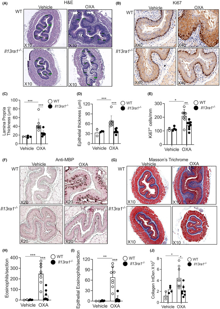
IL‐13Rα1 critically governs EoE pathology. Experimental EoE was induced in wild‐type (WT) and Il13ra1 −/− mice. H&E‐stained slides (A) were analyzed for lamina propria (C) and epithelial (D) thickness. Epithelial cell proliferation was determined using anti‐Ki67 staining (B,E). The slides were stained with anti‐major basic protein (MBP, F), and total (H) and intraepithelial eosinophils were quantified (I). Esophageal fibrosis was determined using Masson's trichrome staining (G,J). Representative photomicrographs of H&E (A), anti‐Ki67 (B), anti‐MBP (F), and Masson's trichrome (G) are shown. Data are representative of n = 3 experiments where each circle represents one mouse. *‐p < 0.05, **‐p < 0.01, ***‐p < 0.001