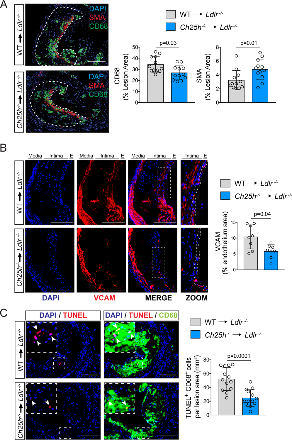
Figure 3. Ch25h deficiency in hematopoietic cells reduces vascular inflammation and promotes plaque stability.

Analysis of Ldlr−/− mice transplanted with WT or Ch25h−/− bone marrow and fed for 12 weeks on a WD. A, Representative histological analysis of cross sections of the aortic sinus stained with CD68 or smooth muscle actin (SMA). Dashed lines delimit the plaque area (A) Quantification of the CD68 or SMA positive area is shown on the right panels and represents the mean ±SD (n=12 mice per group). B, Representative histological analysis of cross sections of the aortic sinus stained with VCAM and DAPI. Quantification of the VCAM positive endothelium area is shown in the right panel and represents the mean ±SD (n=8 mice per group). In the enlarged images the dashed lines show how the endothelium area was delimited for quantification. C, Representative histological analysis of cross sections of the aortic sinus stained with CD68 and TUNEL. Quantification of the CD68-positive cells with TUNEL-positive nuclei is shown in the right panel and represents the mean ±SD (n=13 mice per group). DAPI was used to stain the nuclei. All data were analyzed by Mann-Whitney non-parametric test. Scale bar: 100 μm.
