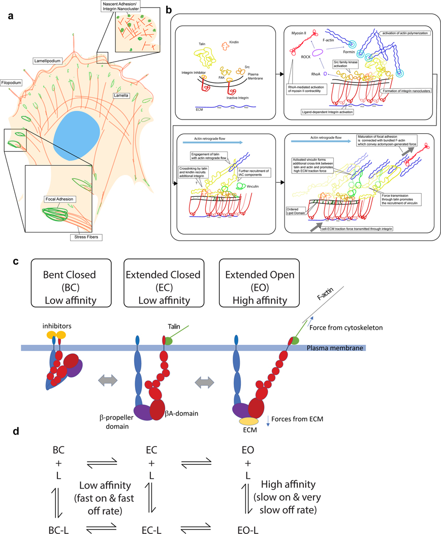
Figure 1 |. Integrin conformational dynamics at integrin-mediated adhesions.
a | Cartoon of the top view of a spread polarised cell with IACs and actin structures indicated. b | Depiction of key steps in the formation and maturation of IACs from initial nascent adhesions to mature FA. c | General schematic depicting the three major conformations of integrin – bent closed (BC), extended closed (EC) and extended open (EO) – along with key interacting proteins and sites of force application. α-subunit is shown in blue and β-subunit in red. Inhibitors (such as filamin199,200, ICAP-1201, or SHARPIN202) binding to α or β cytoplasmic tails are depicted as yellow circles while activating proteins such as talin and kindlin that directly or indirectly link to the cytoskeleton are shown in green. d | Reaction scheme showing the kinetics of integrin and ligand binding taking into account integrin conformational change – based on results in 31.

