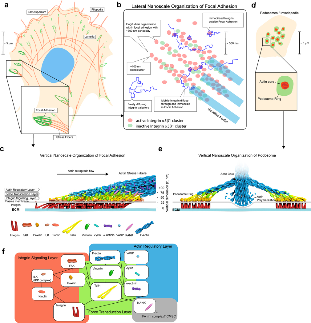
Figure 3 |. Molecular organisation of IACs.
a - b | Lateral composite nanocluster organisation. In mature FAs, extensive granularity and occasional alignments of nanoclusters into linear chains along actin templates can be observed in certain cell types203,204. For integrin α5β1, active and inactive integrin segregate into distinct nanoclusters61 suggesting that nanoclusters could function as discrete units. Single-molecule analysis of integrin motion provides a consistent picture whereby integrin can diffuse freely through IACs. Integrin is immobilised in distinct loci which are comparatively enriched in IACs. c | Multilaminar architecture of FA. Schematic diagram depicting vertical organisation of various IAC components that have been analysed by SRM studies59,87,89,205. Integrins are depicted in clusters of inactive (bent-closed), active (extended-open), as well as in tilted orientation as inferred from fluorescence anisotropy measurements137. d | Podosomes or invadopodia are arrays of typically centrally located IAC structures, consisting of actin-rich core that protrude against and degrade ECM, surrounded by ring-like plaques containing IAC components. e | Vertical organisation of podosome ring featuring polarised orientation of talin, and similar organisation of paxillin and vinculin as in FA. f | Comparison of SRM-based spatial organization with sub-networks of IAC components identified by proteomic analysis (Fig. 2d). A putative IAC compartment anchored by KANK proteins that may potentially interface with microtubules is also depicted.

