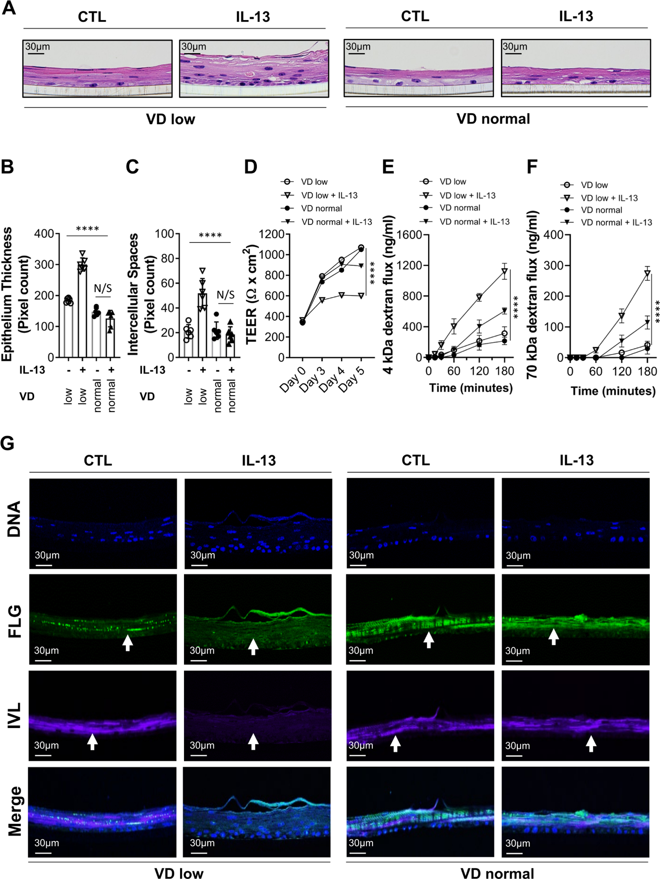
Figure 3.

Effect of vitamin D (VD) on IL-13-dependent epithelial barrier dysfunction. (A–E) The oesophageal epithelial cell air-liquid interface (ALI) organotypic culture was maintained in VD low (0.5 nM) or normal (100 nM) conditions and supplemented with IL-13 (100 ng/mL) or medium alone (control; CTL) for 5 days. (A) ALI cultures were harvested on day 5 and H&E stained. (B–C) Epithelial barrier histopathological quantification of epithelium thickness (B) and intercellular spaces (C) based on H&E staining. (D–F) Epithelial barrier permeability quantification of transepithelial electrical resistance (TEER) (D), 4 kDa FITC-dextran (E) and 70 kDa Texas Red-dextran flux (G) were performed as indicated. (G) Immunofluorescent detection of individual and merged staining of FLG and IVL epithelial structural differentiation markers and cell nuclei (DNA; DAPI). Arrows show marker staining in the ALI culture as indicated. Data are representative (A, G) of n=3 independent experiments or a summary of n=3 (B–D) and n=2 (E, F) independent experiments. Each data point is a mean of a technical duplicate ±SD of individual in vitro assay measurements. Statistics by two-way ANOVA with Tukey’s multiple comparisons test: ****p≤0.0001; ANOVA, analysis of variance; FLG, filaggrin; IVL, involucrin; N/S, not significant.
